Atera WTA Breast Cancer on PDC#
This tutorial records a reproducible PDC run of Agentic Spatial Pathologist on the 10x Xenium Atera WTA Preview FFPE Breast Cancer dataset, using the local pathology-ai backend and pyXenium core workflows.
Dataset#
PDC dataset:
/cfs/klemming/projects/supr/naiss2025-22-606/data/WTA_Preview_FFPE_Breast_Cancer_outsLocal Windows mirror:
Y:\long\10X_datasets\Xenium\Atera\WTA_Preview_FFPE_Breast_Cancer_outsPDC output root:
/cfs/klemming/projects/supr/naiss2025-22-606/results/agentic-spatial-pathologist/atera_wta_breast_pdc_20260429
The dataset copy contains cell_feature_matrix.h5, cell and nucleus boundary parquet files, cells.parquet, experiment.xenium, metrics_summary.csv, WTA_Preview_FFPE_Breast_Cancer_cell_groups.csv, and a registered H&E image pyramid under spatialdata.zarr/images/he.
Service Check#
The workflow uses the PDC local pathology-ai API as the review backend:
curl http://nid002805:8000/health
The completed multimodal run used Slurm job 20163548 on node nid002805. The service job later reached its intended 8-hour test limit, but the workflow artifacts below had already been written. The captured /health payload during the run was:
{
  "service": "pathology-ai",
  "ready": true,
  "settings": {
    "host": "0.0.0.0",
    "port": 8000,
    "llm_base_url": "http://127.0.0.1:8001/v1",
    "llm_model": "openai/gpt-oss-120b",
    "embed_base_url": "http://127.0.0.1:8002",
    "embed_model": "BAAI/bge-m3",
    "rerank_base_url": "http://127.0.0.1:8003",
    "rerank_model": "BAAI/bge-reranker-v2-m3",
    "vector_db": "qdrant",
    "qdrant_base_url": "http://127.0.0.1:6333",
    "qdrant_collection": "pathology_reference",
    "default_top_k": 6,
    "strict_json": true,
    "max_chunk_chars": 1200,
    "chunk_overlap_chars": 120
  },
  "components": {
    "llm": {
      "ok": true,
      "models": [
        "openai/gpt-oss-120b"
      ]
    },
    "embedder": {
      "ok": true
    },
    "reranker": {
      "ok": true
    },
    "vector_store": {
      "ok": true,
      "collections": []
    }
  }
}
Submit the PDC Job#
From the PDC login node:
cd /cfs/klemming/home/h/hutaobo/Agentic-Spatial-Pathologist
git fetch origin
git checkout main
git pull --ff-only origin main

sbatch \
  --export=ALL,PATHOLOGY_AI_BASE_URL=http://nid002805:8000 \
  deploy/pathology_ai/atera_wta_breast_pdc.sbatch
The Slurm wrapper creates a venv under the output root, installs spatho, pyXenium, histoseg, and scientific dependencies, clones sfplot for tissue_structure_pipeline, then runs:
python scripts/pdc_atera_breast_workflow.py \
  --dataset-root /cfs/klemming/projects/supr/naiss2025-22-606/data/WTA_Preview_FFPE_Breast_Cancer_outs \
  --run-root /cfs/klemming/projects/supr/naiss2025-22-606/results/agentic-spatial-pathologist/atera_wta_breast_pdc_20260429 \
  --sfplot-root /cfs/klemming/projects/supr/naiss2025-22-606/results/agentic-spatial-pathologist/atera_wta_breast_pdc_20260429/deps/sfplot \
  --pathology-ai-base-url http://nid002805:8000
Generated Inputs#
The source dataset already has graph-cluster assignments, but it does not include the standard 10x differential-expression and UMAP projection CSVs expected by the full-auto spatho workflow. The PDC driver generates:
inputs/analysis/clustering/gene_expression_graphclust/clusters.csvinputs/analysis/diffexp/gene_expression_graphclust/differential_expression.csvinputs/analysis/umap/gene_expression_2_components/projection.csvinputs/he/atera_wta_breast_pdc_20260429_registered_he_level6.tifinputs/he/atera_wta_breast_pdc_20260429_he_alignment_level6.csv
The differential-expression table is a cluster-pseudobulk log2 fold-change approximation from cell_feature_matrix.h5; the projection is derived from cell centroids. The H&E tutorial asset is extracted from spatialdata.zarr/images/he level 6 and uses the stored affine transform from image pixel coordinates to Xenium pixel coordinates.
{
  "cluster_source": "/cfs/klemming/projects/supr/naiss2025-22-606/data/WTA_Preview_FFPE_Breast_Cancer_outs/analysis/analysis/clustering/gene_expression_graphclust/clusters.csv",
  "cluster_csv": "/cfs/klemming/projects/supr/naiss2025-22-606/results/ai-driven-spatial-pathologist/atera_wta_breast_pdc_20260429/inputs/analysis/clustering/gene_expression_graphclust/clusters.csv",
  "projection": {
    "projection_csv": "/cfs/klemming/projects/supr/naiss2025-22-606/results/ai-driven-spatial-pathologist/atera_wta_breast_pdc_20260429/inputs/analysis/umap/gene_expression_2_components/projection.csv",
    "source": "cell_centroids",
    "rows": 170045,
    "matched_fraction": 1.0
  },
  "differential_expression": {
    "differential_expression_csv": "/cfs/klemming/projects/supr/naiss2025-22-606/results/ai-driven-spatial-pathologist/atera_wta_breast_pdc_20260429/inputs/analysis/diffexp/gene_expression_graphclust/differential_expression.csv",
    "genes": 27104,
    "clusters": [
      "1",
      "2",
      "3",
      "4",
      "5",
      "6",
      "7",
      "8",
      "9",
      "10",
      "11",
      "12",
      "13",
      "14",
      "15",
      "16",
      "17",
      "18",
      "19",
      "20",
      "21",
      "22",
      "23",
      "24",
      "25",
      "26",
      "27",
      "28",
      "29",
      "30",
      "31",
      "32",
      "33",
      "34"
    ],
    "matched_barcodes": 170045,
    "total_barcodes": 170057,
    "method": "cluster_pseudobulk_log2fc_with_thresholded_p_values"
  }
}
Agentic Spatial Pathologist Run#
The current workflow config uses the local backend and keeps OpenAI disabled:
{
  "annotation_taxonomy": "breast",
  "pathology_review_backend": "pathology_ai_api",
  "pathology_ai_api_base_url": "http://nid002805:8000",
  "cluster_annotation_backend": "pathology_ai_api",
  "cluster_annotation_llm_base_url": "http://nid002805:8000",
  "he_contour_foundation_enabled": true,
  "he_foundation_model_id": "vinid/plip",
  "he_foundation_prompt_set": "breast_contour_v1",
  "he_visual_override_enabled": true,
  "rna_foundation_enabled": true,
  "rna_foundation_backend": "precomputed_scgpt",
  "rna_foundation_cell_mapping_path": "inputs/foundation/scgpt_cell_mapping.csv",
  "pathway_activity_enabled": true,
  "pathway_activity_csv": null,
  "niche_fusion_enabled": true,
  "niche_fusion_backend": "lightweight",
  "openai_enabled": false
}
With cluster_annotation_backend="pathology_ai_api", reruns write cluster_celltype_annotation.csv as a conservative consensus: marker-based heuristic labels are reviewed by the local PDC LLM, accepted only when the local model returns a valid controlled-vocabulary label with enough confidence and marker support. The paid OpenAI API is not used.
With he_contour_foundation_enabled=true, the workflow follows the pyXenium RNA + contour + H&E pattern: it imports xenium_explorer_annotations.generated.geojson, extracts aligned masked H&E contour patches from spatialdata.zarr/images/he, classifies those patches locally through PLIP (vinid/plip), and asks the local PDC LLM to fuse visual evidence with RNA/cell-type structure evidence for final structure names. Accepted visual overrides are recorded explicitly rather than silently replacing the molecular-only labels.
The readiness check is:
spatho doctor --config workflows/atera_wta_breast_pdc_20260429_pathology_ai.json
The captured doctor output and workflow summary are included here:
{
  "openai_api_key_present": false,
  "openai_api_key_length": 0,
  "config_path": "/cfs/klemming/projects/supr/naiss2025-22-606/results/ai-driven-spatial-pathologist/atera_wta_breast_pdc_20260429/workflows/atera_wta_breast_pdc_20260429_pathology_ai.json",
  "config_exists": true,
  "case_name": "atera_wta_breast_pdc_20260429",
  "annotation_taxonomy": "breast",
  "pathology_review_backend": "pathology_ai_api",
  "pathology_ai_api_base_url": "http://127.0.0.1:8000",
  "pathology_ai_top_k": 6,
  "pathology_ai_answer_language": "en",
  "pathology_ai_document_ids": [],
  "cluster_annotation_backend": "pathology_ai_api",
  "cluster_annotation_llm_base_url": "http://127.0.0.1:8000",
  "cluster_annotation_min_llm_confidence": 0.6,
  "cluster_annotation_override_margin": 0.15,
  "cluster_annotation_require_marker_overlap": true,
  "he_contour_foundation_enabled": true,
  "he_contour_geojson": "/cfs/klemming/projects/supr/naiss2025-22-606/data/WTA_Preview_FFPE_Breast_Cancer_outs/xenium_explorer_annotations.generated.geojson",
  "he_contour_key": "atera_wta_breast_he_contours",
  "he_foundation_model_id": "vinid/plip",
  "he_foundation_prompt_set": "breast_contour_v1",
  "he_foundation_top_k": 5,
  "he_foundation_max_patch_side_px": 1024,
  "he_visual_override_enabled": true,
  "he_visual_override_min_llm_confidence": 0.7,
  "he_visual_override_min_foundation_score": 0.35,
  "schema_valid": true,
  "organ_pack": {
    "id": "breast",
    "display_name": "Breast Cancer Xenium",
    "annotation_taxonomy": "breast",
    "description": "Built-in organ pack for breast Xenium cases with breast-specific cluster annotation defaults, public workflow template defaults, and expected artifact groups.",
    "default_study_context": "Human breast cancer Xenium case. Run breast-specific OpenAI-driven cluster annotation from graph-based differential expression evidence, then propagate the generated labels through structure discovery, H&E overlay, and AI spatial pathology review. Treat this as pathology decision support, not an autonomous diagnosis.",
    "supported_input_layout": "Xenium outs directory with analysis/diffexp/gene_expression_graphclust and analysis/umap/gene_expression_2_components.",
    "workflow_defaults": {
      "top_positive_markers": 15,
      "top_negative_markers": 6,
      "min_log2fc": 0.5,
      "max_adjusted_p_value": 0.05,
      "top_neighbors": 5,
      "low_confidence_threshold": 0.65,
      "ambiguity_margin_threshold": 0.08,
      "top_clusters_per_structure": 8,
      "openai_model": "gpt-5.4",
      "openai_reasoning_effort": "medium",
      "openai_store": false
    },
    "artifact_contract": {
      "required": [
        "annotation.cluster_evidence_json",
        "annotation.cluster_annotations_json",
        "annotation.compatibility_csv",
        "annotation.report_html",
        "pathology.report_html",
        "pathology.structure_reviews_json",
        "pathology.case_summary_json",
        "pipeline.structure_clustermap_pdf",
        "pipeline.cluster_structure_lookup_csv",
        "workflow.workflow_summary_json"
      ]
    }
  },
  "base_pipeline_config": "/cfs/klemming/projects/supr/naiss2025-22-606/results/ai-driven-spatial-pathologist/atera_wta_breast_pdc_20260429/runtime/segmentation_methods/projects/atera_wta_breast_pdc_20260429/configs/atera_wta_breast_pdc_20260429_base_pipeline.json",
  "base_pipeline_config_exists": true,
  "differential_expression_csv": "/cfs/klemming/projects/supr/naiss2025-22-606/results/ai-driven-spatial-pathologist/atera_wta_breast_pdc_20260429/inputs/analysis/diffexp/gene_expression_graphclust/differential_expression.csv",
  "differential_expression_csv_exists": true,
  "projection_csv": "/cfs/klemming/projects/supr/naiss2025-22-606/results/ai-driven-spatial-pathologist/atera_wta_breast_pdc_20260429/inputs/analysis/umap/gene_expression_2_components/projection.csv",
  "projection_csv_exists": true,
  "output_root": "/cfs/klemming/projects/supr/naiss2025-22-606/results/ai-driven-spatial-pathologist/atera_wta_breast_pdc_20260429/runtime/segmentation_methods/projects/atera_wta_breast_pdc_20260429/outputs/spatho",
  "dataset_modality": "xenium_rna_protein",
  "canonical_space": "physical_um",
  "export_space": "xenium_explorer_pixel",
  "xenium_pixel_size_um": 0.2125,
  "segmentation_source": "ranger_default",
  "openai_enabled": false,
  "openai_model": "gpt-5.4",
  "pathology_ai_api_health": {
    "service": "pathology-ai",
    "ready": true,
    "settings": {
      "host": "0.0.0.0",
      "port": 8000,
      "llm_base_url": "http://127.0.0.1:8001/v1",
      "llm_model": "openai/gpt-oss-120b",
      "embed_base_url": "http://127.0.0.1:8002",
      "embed_model": "BAAI/bge-m3",
      "rerank_base_url": "http://127.0.0.1:8003",
      "rerank_model": "BAAI/bge-reranker-v2-m3",
      "vector_db": "qdrant",
      "qdrant_base_url": "http://127.0.0.1:6333",
      "qdrant_collection": "pathology_reference",
      "default_top_k": 6,
      "strict_json": true,
      "max_chunk_chars": 1200,
      "chunk_overlap_chars": 120
    },
    "components": {
      "llm": {
        "ok": true,
        "models": [
          "openai/gpt-oss-120b"
        ]
      },
      "embedder": {
        "ok": true
      },
      "reranker": {
        "ok": true
      },
      "vector_store": {
        "ok": true,
        "collections": []
      }
    }
  },
  "cluster_annotation_api_health": {
    "service": "pathology-ai",
    "ready": true,
    "settings": {
      "host": "0.0.0.0",
      "port": 8000,
      "llm_base_url": "http://127.0.0.1:8001/v1",
      "llm_model": "openai/gpt-oss-120b",
      "embed_base_url": "http://127.0.0.1:8002",
      "embed_model": "BAAI/bge-m3",
      "rerank_base_url": "http://127.0.0.1:8003",
      "rerank_model": "BAAI/bge-reranker-v2-m3",
      "vector_db": "qdrant",
      "qdrant_base_url": "http://127.0.0.1:6333",
      "qdrant_collection": "pathology_reference",
      "default_top_k": 6,
      "strict_json": true,
      "max_chunk_chars": 1200,
      "chunk_overlap_chars": 120
    },
    "components": {
      "llm": {
        "ok": true,
        "models": [
          "openai/gpt-oss-120b"
        ]
      },
      "embedder": {
        "ok": true
      },
      "reranker": {
        "ok": true
      },
      "vector_store": {
        "ok": true,
        "collections": []
      }
    }
  },
  "he_contour_geojson_resolved": "/cfs/klemming/projects/supr/naiss2025-22-606/data/WTA_Preview_FFPE_Breast_Cancer_outs/xenium_explorer_annotations.generated.geojson",
  "he_foundation_api_health": {
    "service": "pathology-ai",
    "ready": true,
    "settings": {
      "host": "0.0.0.0",
      "port": 8000,
      "llm_base_url": "http://127.0.0.1:8001/v1",
      "llm_model": "openai/gpt-oss-120b",
      "embed_base_url": "http://127.0.0.1:8002",
      "embed_model": "BAAI/bge-m3",
      "rerank_base_url": "http://127.0.0.1:8003",
      "rerank_model": "BAAI/bge-reranker-v2-m3",
      "vector_db": "qdrant",
      "qdrant_base_url": "http://127.0.0.1:6333",
      "qdrant_collection": "pathology_reference",
      "default_top_k": 6,
      "strict_json": true,
      "max_chunk_chars": 1200,
      "chunk_overlap_chars": 120
    },
    "components": {
      "llm": {
        "ok": true,
        "models": [
          "openai/gpt-oss-120b"
        ]
      },
      "embedder": {
        "ok": true
      },
      "reranker": {
        "ok": true
      },
      "vector_store": {
        "ok": true,
        "collections": []
      }
    }
  },
  "issues": [],
  "ready_to_run": true
}
{
  "case_name": "atera_wta_breast_pdc_20260429",
  "study_context": "10x Xenium WTA Preview FFPE Breast Cancer sample on PDC. Graph-based clusters are annotated with breast taxonomy, reviewed through the local pathology-ai API, and paired with pyXenium topology and mechanostress outputs.",
  "output_root": "/cfs/klemming/projects/supr/naiss2025-22-606/results/ai-driven-spatial-pathologist/atera_wta_breast_pdc_20260429/runtime/segmentation_methods/projects/atera_wta_breast_pdc_20260429/outputs/spatho",
  "annotation_outputs": {
    "output_dir": "/cfs/klemming/projects/supr/naiss2025-22-606/results/ai-driven-spatial-pathologist/atera_wta_breast_pdc_20260429/runtime/segmentation_methods/projects/atera_wta_breast_pdc_20260429/outputs/spatho/annotation",
    "cluster_evidence_json": "/cfs/klemming/projects/supr/naiss2025-22-606/results/ai-driven-spatial-pathologist/atera_wta_breast_pdc_20260429/runtime/segmentation_methods/projects/atera_wta_breast_pdc_20260429/outputs/spatho/annotation/cluster_evidence.json",
    "cluster_annotations_json": "/cfs/klemming/projects/supr/naiss2025-22-606/results/ai-driven-spatial-pathologist/atera_wta_breast_pdc_20260429/runtime/segmentation_methods/projects/atera_wta_breast_pdc_20260429/outputs/spatho/annotation/cluster_annotations_openai.json",
    "cluster_annotations_csv": "/cfs/klemming/projects/supr/naiss2025-22-606/results/ai-driven-spatial-pathologist/atera_wta_breast_pdc_20260429/runtime/segmentation_methods/projects/atera_wta_breast_pdc_20260429/outputs/spatho/annotation/cluster_annotations_openai.csv",
    "compatibility_csv": "/cfs/klemming/projects/supr/naiss2025-22-606/results/ai-driven-spatial-pathologist/atera_wta_breast_pdc_20260429/runtime/segmentation_methods/projects/atera_wta_breast_pdc_20260429/outputs/spatho/annotation/cluster_celltype_annotation.csv",
    "case_review_json": "/cfs/klemming/projects/supr/naiss2025-22-606/results/ai-driven-spatial-pathologist/atera_wta_breast_pdc_20260429/runtime/segmentation_methods/projects/atera_wta_breast_pdc_20260429/outputs/spatho/annotation/annotation_case_review.json",
    "report_html": "/cfs/klemming/projects/supr/naiss2025-22-606/results/ai-driven-spatial-pathologist/atera_wta_breast_pdc_20260429/runtime/segmentation_methods/projects/atera_wta_breast_pdc_20260429/outputs/spatho/annotation/annotation_review.html",
    "heuristic_annotations_json": "/cfs/klemming/projects/supr/naiss2025-22-606/results/ai-driven-spatial-pathologist/atera_wta_breast_pdc_20260429/runtime/segmentation_methods/projects/atera_wta_breast_pdc_20260429/outputs/spatho/annotation/cluster_annotations_heuristic.json",
    "heuristic_annotations_csv": "/cfs/klemming/projects/supr/naiss2025-22-606/results/ai-driven-spatial-pathologist/atera_wta_breast_pdc_20260429/runtime/segmentation_methods/projects/atera_wta_breast_pdc_20260429/outputs/spatho/annotation/cluster_annotations_heuristic.csv",
    "pathology_ai_annotations_json": "/cfs/klemming/projects/supr/naiss2025-22-606/results/ai-driven-spatial-pathologist/atera_wta_breast_pdc_20260429/runtime/segmentation_methods/projects/atera_wta_breast_pdc_20260429/outputs/spatho/annotation/cluster_annotations_pathology_ai.json",
    "pathology_ai_annotations_csv": "/cfs/klemming/projects/supr/naiss2025-22-606/results/ai-driven-spatial-pathologist/atera_wta_breast_pdc_20260429/runtime/segmentation_methods/projects/atera_wta_breast_pdc_20260429/outputs/spatho/annotation/cluster_annotations_pathology_ai.csv",
    "consensus_annotations_json": "/cfs/klemming/projects/supr/naiss2025-22-606/results/ai-driven-spatial-pathologist/atera_wta_breast_pdc_20260429/runtime/segmentation_methods/projects/atera_wta_breast_pdc_20260429/outputs/spatho/annotation/cluster_annotations_consensus.json",
    "consensus_annotations_csv": "/cfs/klemming/projects/supr/naiss2025-22-606/results/ai-driven-spatial-pathologist/atera_wta_breast_pdc_20260429/runtime/segmentation_methods/projects/atera_wta_breast_pdc_20260429/outputs/spatho/annotation/cluster_annotations_consensus.csv",
    "annotation_refinement_metadata_json": "/cfs/klemming/projects/supr/naiss2025-22-606/results/ai-driven-spatial-pathologist/atera_wta_breast_pdc_20260429/runtime/segmentation_methods/projects/atera_wta_breast_pdc_20260429/outputs/spatho/annotation/annotation_refinement_metadata.json"
  },
  "runtime_base_pipeline_config": "/cfs/klemming/projects/supr/naiss2025-22-606/results/ai-driven-spatial-pathologist/atera_wta_breast_pdc_20260429/runtime/segmentation_methods/projects/atera_wta_breast_pdc_20260429/outputs/spatho/runtime_configs/generated_runtime_config.json",
  "pathology_outputs": {
    "output_dir": "/cfs/klemming/projects/supr/naiss2025-22-606/results/ai-driven-spatial-pathologist/atera_wta_breast_pdc_20260429/runtime/segmentation_methods/projects/atera_wta_breast_pdc_20260429/outputs/spatho/pathology_review",
    "report_html": "/cfs/klemming/projects/supr/naiss2025-22-606/results/ai-driven-spatial-pathologist/atera_wta_breast_pdc_20260429/runtime/segmentation_methods/projects/atera_wta_breast_pdc_20260429/outputs/spatho/pathology_review/index.html",
    "cluster_reviews_json": "/cfs/klemming/projects/supr/naiss2025-22-606/results/ai-driven-spatial-pathologist/atera_wta_breast_pdc_20260429/runtime/segmentation_methods/projects/atera_wta_breast_pdc_20260429/outputs/spatho/pathology_review/cluster_reviews.json",
    "structure_reviews_json": "/cfs/klemming/projects/supr/naiss2025-22-606/results/ai-driven-spatial-pathologist/atera_wta_breast_pdc_20260429/runtime/segmentation_methods/projects/atera_wta_breast_pdc_20260429/outputs/spatho/pathology_review/structure_reviews.json",
    "case_summary_json": "/cfs/klemming/projects/supr/naiss2025-22-606/results/ai-driven-spatial-pathologist/atera_wta_breast_pdc_20260429/runtime/segmentation_methods/projects/atera_wta_breast_pdc_20260429/outputs/spatho/pathology_review/case_summary.json"
  },
  "responses_api_ready": true,
  "annotation_taxonomy": "breast",
  "cluster_annotation_backend": "pathology_ai_api",
  "cluster_annotation_llm_base_url": "http://127.0.0.1:8000",
  "cluster_annotation_min_llm_confidence": 0.6,
  "cluster_annotation_override_margin": 0.15,
  "cluster_annotation_require_marker_overlap": true,
  "pathology_review_backend": "pathology_ai_api",
  "pathology_ai_api_base_url": "http://127.0.0.1:8000",
  "pathology_ai_top_k": 6,
  "pathology_ai_answer_language": "en",
  "pathology_ai_document_ids": [],
  "pipeline_recomputed": true,
  "openai_enabled": false,
  "openai_api_key_env": "OPENAI_API_KEY",
  "openai_model": "gpt-5.4",
  "openai_reasoning_effort": "medium",
  "openai_store": false,
  "he_foundation_outputs": {
    "he_foundation_dir": "/cfs/klemming/projects/supr/naiss2025-22-606/results/ai-driven-spatial-pathologist/atera_wta_breast_pdc_20260429/runtime/segmentation_methods/projects/atera_wta_breast_pdc_20260429/outputs/spatho/he_foundation",
    "patch_manifest_json": "/cfs/klemming/projects/supr/naiss2025-22-606/results/ai-driven-spatial-pathologist/atera_wta_breast_pdc_20260429/runtime/segmentation_methods/projects/atera_wta_breast_pdc_20260429/outputs/spatho/he_foundation/he_contour_patches_manifest.json",
    "classification_json": "/cfs/klemming/projects/supr/naiss2025-22-606/results/ai-driven-spatial-pathologist/atera_wta_breast_pdc_20260429/runtime/segmentation_methods/projects/atera_wta_breast_pdc_20260429/outputs/spatho/he_foundation/he_contour_classification.json",
    "classification_csv": "/cfs/klemming/projects/supr/naiss2025-22-606/results/ai-driven-spatial-pathologist/atera_wta_breast_pdc_20260429/runtime/segmentation_methods/projects/atera_wta_breast_pdc_20260429/outputs/spatho/he_foundation/he_contour_classification.csv",
    "structure_summary_json": "/cfs/klemming/projects/supr/naiss2025-22-606/results/ai-driven-spatial-pathologist/atera_wta_breast_pdc_20260429/runtime/segmentation_methods/projects/atera_wta_breast_pdc_20260429/outputs/spatho/he_foundation/he_contour_to_structure_summary.json",
    "structure_summary_csv": "/cfs/klemming/projects/supr/naiss2025-22-606/results/ai-driven-spatial-pathologist/atera_wta_breast_pdc_20260429/runtime/segmentation_methods/projects/atera_wta_breast_pdc_20260429/outputs/spatho/he_foundation/he_contour_to_structure_summary.csv",
    "structure_multimodal_names_json": "/cfs/klemming/projects/supr/naiss2025-22-606/results/ai-driven-spatial-pathologist/atera_wta_breast_pdc_20260429/runtime/segmentation_methods/projects/atera_wta_breast_pdc_20260429/outputs/spatho/he_foundation/structure_multimodal_names.json",
    "structure_multimodal_names_csv": "/cfs/klemming/projects/supr/naiss2025-22-606/results/ai-driven-spatial-pathologist/atera_wta_breast_pdc_20260429/runtime/segmentation_methods/projects/atera_wta_breast_pdc_20260429/outputs/spatho/he_foundation/structure_multimodal_names.csv",
    "updated_structure_reviews_json": "/cfs/klemming/projects/supr/naiss2025-22-606/results/ai-driven-spatial-pathologist/atera_wta_breast_pdc_20260429/runtime/segmentation_methods/projects/atera_wta_breast_pdc_20260429/outputs/spatho/pathology_review/structure_reviews.json",
    "updated_structure_reviews_csv": "/cfs/klemming/projects/supr/naiss2025-22-606/results/ai-driven-spatial-pathologist/atera_wta_breast_pdc_20260429/runtime/segmentation_methods/projects/atera_wta_breast_pdc_20260429/outputs/spatho/pathology_review/structure_reviews.csv",
    "updated_case_summary_json": "/cfs/klemming/projects/supr/naiss2025-22-606/results/ai-driven-spatial-pathologist/atera_wta_breast_pdc_20260429/runtime/segmentation_methods/projects/atera_wta_breast_pdc_20260429/outputs/spatho/pathology_review/case_summary.json",
    "updated_report_html": "/cfs/klemming/projects/supr/naiss2025-22-606/results/ai-driven-spatial-pathologist/atera_wta_breast_pdc_20260429/runtime/segmentation_methods/projects/atera_wta_breast_pdc_20260429/outputs/spatho/pathology_review/index.html",
    "accepted_visual_overrides": "0",
    "metadata_json": "/cfs/klemming/projects/supr/naiss2025-22-606/results/ai-driven-spatial-pathologist/atera_wta_breast_pdc_20260429/runtime/segmentation_methods/projects/atera_wta_breast_pdc_20260429/outputs/spatho/he_foundation/he_foundation_metadata.json"
  }
}
scGPT/SpatialFusion-Inspired Evidence Layer#
The upgraded workflow can add a foundation evidence layer between cluster annotation and structure-level pathology review. This layer is opt-in and does not change the OpenAI API path or the local pathology-ai path.
For this Atera breast run, the intended PDC configuration is:
rna_foundation_enabled=truewhen a precomputed scGPT/scGPT-spatial cell mapping is available;pathway_activity_enabled=true, using the generated differential-expression CSV when no pathway activity table is supplied;niche_fusion_enabled=true, using lightweight fusion of RNA reference evidence, pathway scores, PLIP H&E morphology signals, and spatial structure metadata.
If a real scGPT/scGPT-spatial mapping has not been generated yet, the PDC tutorial driver writes a zero-confidence smoke mapping at inputs/foundation/scgpt_cell_mapping.csv. That file validates the data interface and report plumbing only; replace it with a real reference-mapping table before interpreting RNA foundation labels biologically.
The layer writes:
foundation/rna_foundation_cluster_summary.csvfoundation/rna_foundation_structure_summary.csvfoundation/pathway_activity_structure_summary.csvfoundation/he_morphology_feature_summary.csvfoundation/niche_fusion_summary.csvfoundation/foundation_evidence_metadata.json
The method is inspired by scGPT and SpatialFusion, but it remains an auditable workflow layer: frozen/precomputed RNA evidence and H&E foundation-model scores are summarized into standard tables, then the local LLM records whether modalities agree, conflict, or complement each other. It does not train a new joint embedding model in this tutorial pass.
Interpretation policy:
RNA/cell-type and marker evidence preserve the primary biological identity of each structure.
PLIP H&E contour evidence contributes morphology, inflammation, tumor, stroma, and artifact signals.
Pathway activity contributes molecular program context, for example epithelial tumor, proliferation, stromal, immune, or hypoxia programs.
The local LLM adjudicates consistency: visual evidence can support, qualify, or challenge a label, but conservative thresholds prevent silent replacement.
Selected overlays:

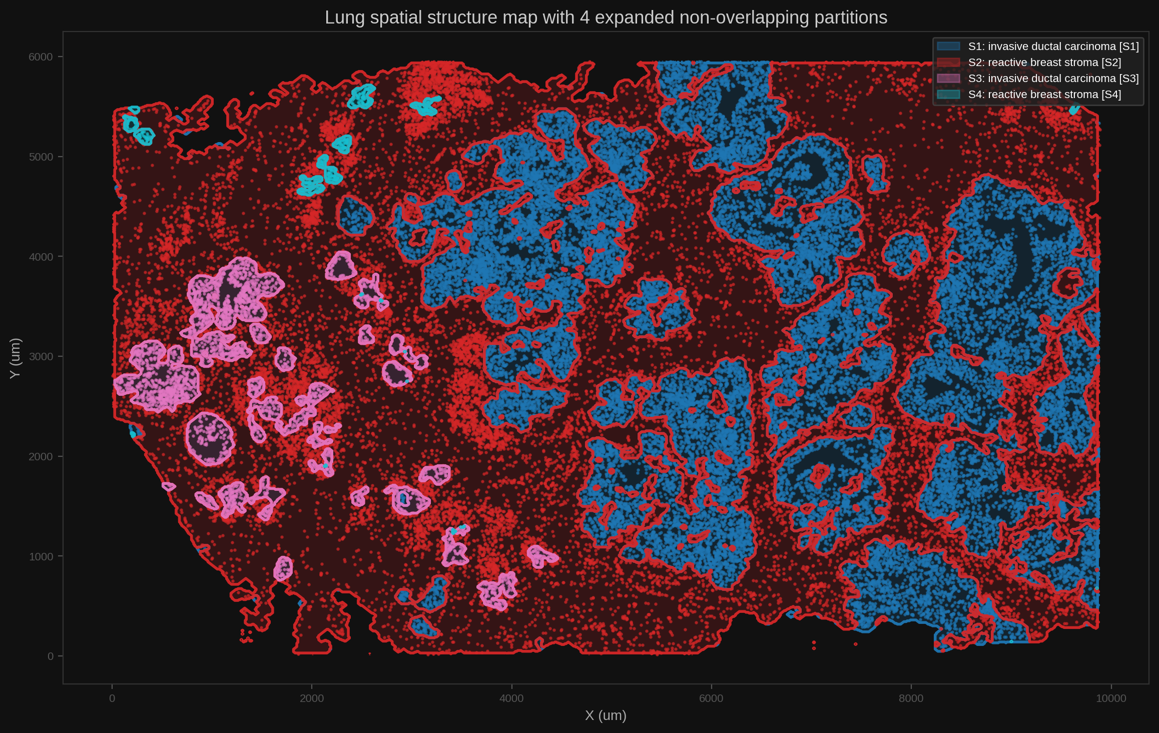
Local LLM + H&E Foundation Results#
The real PDC run classified 2606 aligned H&E contour patches with the local PLIP pathology foundation model (vinid/plip) and wrote 2606 contour-level predictions. Those patch-level predictions were aggregated into 7 structure-level visual summaries. The local LLM then fused the PLIP visual evidence with RNA/cell-type/structure evidence for 4 downstream structure names.
The key point is that PLIP is used as local visual evidence, not as an automatic label replacement. The local LLM checks whether the visual foundation-model signal agrees with, contradicts, or complements the molecular interpretation. In this run accepted_visual_overrides=0, which is the intended conservative behavior: no H&E-only signal was strong enough, and consistent enough with RNA/cell-type evidence, to replace the molecularly supported IDC or stroma labels.
{
  "enabled": true,
  "contour_geojson": "/cfs/klemming/projects/supr/naiss2025-22-606/data/WTA_Preview_FFPE_Breast_Cancer_outs/xenium_explorer_annotations.generated.geojson",
  "contour_key": "atera_wta_breast_he_contours",
  "model_id": "vinid/plip",
  "prompt_set": "breast_contour_v1",
  "top_k": 5,
  "max_patch_side_px": 1024,
  "patch_count": 2606,
  "classification_count": 2606,
  "classification_warnings": [],
  "visual_override_enabled": true,
  "visual_override_min_llm_confidence": 0.7,
  "visual_override_min_foundation_score": 0.35,
  "multimodal_naming_rerun_base_url": "http://127.0.0.1:8010",
  "multimodal_naming_rerun_reason": "normalize local LLM evidence lists"
}
Structure-level PLIP aggregation:
structure_id,structure_name,n_contours,n_classified,top_label_id,top_label,top_mean_score,top_max_score,top_visual_labels_json
1,11q13 Invasive Tumor Cells,136,136,artifact_low_quality,Artifact or low-quality tissue,0.34652924849424604,0.8685594797134399,"[{""label_id"": ""artifact_low_quality"", ""label"": ""Artifact or low-quality tissue"", ""contour_count"": 82, ""mean_score"": 0.34652924849424604, ""max_score"": 0.8685594797134399}, {""label_id"": ""macrophage_inflammation"", ""label"": ""Macrophage-rich inflammation"", ""contour_count"": 131, ""mean_score"": 0.32548482953535235, ""max_score"": 0.9525430798530579}, {""label_id"": ""dcis_or_ductal_tumor"", ""label"": ""Ductal carcinoma in situ or ductal tumor"", ""contour_count"": 54, ""mean_score"": 0.1819703367245556, ""max_score"": 0.5444559454917908}, {""label_id"": ""invasive_tumor_epithelium"", ""label"": ""Invasive tumor epithelium"", ""contour_count"": 57, ""mean_score"": 0.17000476189219116, ""max_score"": 0.5119240283966064}, {""label_id"": ""adipose_or_empty"", ""label"": ""Adipose or low-cellularity tissue"", ""contour_count"": 72, ""mean_score"": 0.15550945124899349, ""max_score"": 0.45895981788635254}]"
2,Basal-like Structured DCIS Cells,548,548,macrophage_inflammation,Macrophage-rich inflammation,0.32113183564109393,0.9507338404655457,"[{""label_id"": ""macrophage_inflammation"", ""label"": ""Macrophage-rich inflammation"", ""contour_count"": 516, ""mean_score"": 0.32113183564109393, ""max_score"": 0.9507338404655457}, {""label_id"": ""artifact_low_quality"", ""label"": ""Artifact or low-quality tissue"", ""contour_count"": 452, ""mean_score"": 0.2935638413476074, ""max_score"": 0.8591423034667969}, {""label_id"": ""invasive_tumor_epithelium"", ""label"": ""Invasive tumor epithelium"", ""contour_count"": 129, ""mean_score"": 0.14171397872269154, ""max_score"": 0.49712949991226196}, {""label_id"": ""adipose_or_empty"", ""label"": ""Adipose or low-cellularity tissue"", ""contour_count"": 336, ""mean_score"": 0.14060028432729832, ""max_score"": 0.46402403712272644}, {""label_id"": ""vascular_region"", ""label"": ""Vascular or endothelial region"", ""contour_count"": 391, ""mean_score"": 0.12796525352293878, ""max_score"": 0.6157298684120178}]"
3,Macrophages,695,695,macrophage_inflammation,Macrophage-rich inflammation,0.31556032766793535,0.9596852660179138,"[{""label_id"": ""macrophage_inflammation"", ""label"": ""Macrophage-rich inflammation"", ""contour_count"": 639, ""mean_score"": 0.31556032766793535, ""max_score"": 0.9596852660179138}, {""label_id"": ""artifact_low_quality"", ""label"": ""Artifact or low-quality tissue"", ""contour_count"": 561, ""mean_score"": 0.2320593531673472, ""max_score"": 0.857221782207489}, {""label_id"": ""vascular_region"", ""label"": ""Vascular or endothelial region"", ""contour_count"": 539, ""mean_score"": 0.1988691238035035, ""max_score"": 0.7634395360946655}, {""label_id"": ""fibrocollagenous_stroma"", ""label"": ""Fibrocollagenous or desmoplastic stroma"", ""contour_count"": 429, ""mean_score"": 0.17781993087233502, ""max_score"": 0.9401921629905701}, {""label_id"": ""adipose_or_empty"", ""label"": ""Adipose or low-cellularity tissue"", ""contour_count"": 406, ""mean_score"": 0.14190508644991173, ""max_score"": 0.7174577713012695}]"
4,Plasma Cells,353,353,macrophage_inflammation,Macrophage-rich inflammation,0.4486155729915652,0.9617540240287781,"[{""label_id"": ""macrophage_inflammation"", ""label"": ""Macrophage-rich inflammation"", ""contour_count"": 341, ""mean_score"": 0.4486155729915652, ""max_score"": 0.9617540240287781}, {""label_id"": ""vascular_region"", ""label"": ""Vascular or endothelial region"", ""contour_count"": 333, ""mean_score"": 0.2068247582655758, ""max_score"": 0.8306549191474915}, {""label_id"": ""fibrocollagenous_stroma"", ""label"": ""Fibrocollagenous or desmoplastic stroma"", ""contour_count"": 158, ""mean_score"": 0.1407409678802743, ""max_score"": 0.7920061945915222}, {""label_id"": ""artifact_low_quality"", ""label"": ""Artifact or low-quality tissue"", ""contour_count"": 275, ""mean_score"": 0.13010974021374502, ""max_score"": 0.7871575951576233}, {""label_id"": ""necrosis_debris"", ""label"": ""Necrosis or debris"", ""contour_count"": 324, ""mean_score"": 0.10199108183740374, ""max_score"": 0.5879630446434021}]"
5,Endothelial Cells,735,735,macrophage_inflammation,Macrophage-rich inflammation,0.286686747278517,0.9435091614723206,"[{""label_id"": ""macrophage_inflammation"", ""label"": ""Macrophage-rich inflammation"", ""contour_count"": 669, ""mean_score"": 0.286686747278517, ""max_score"": 0.9435091614723206}, {""label_id"": ""artifact_low_quality"", ""label"": ""Artifact or low-quality tissue"", ""contour_count"": 529, ""mean_score"": 0.2491795973295948, ""max_score"": 0.7932630181312561}, {""label_id"": ""fibrocollagenous_stroma"", ""label"": ""Fibrocollagenous or desmoplastic stroma"", ""contour_count"": 491, ""mean_score"": 0.21493188439168476, ""max_score"": 0.9354937672615051}, {""label_id"": ""vascular_region"", ""label"": ""Vascular or endothelial region"", ""contour_count"": 579, ""mean_score"": 0.1881815644659887, ""max_score"": 0.7514576315879822}, {""label_id"": ""adipose_or_empty"", ""label"": ""Adipose or low-cellularity tissue"", ""contour_count"": 488, ""mean_score"": 0.1370952495938686, ""max_score"": 0.4735677242279053}]"
6,Apocrine Cells,18,18,artifact_low_quality,Artifact or low-quality tissue,0.32160715758800507,0.6824164390563965,"[{""label_id"": ""artifact_low_quality"", ""label"": ""Artifact or low-quality tissue"", ""contour_count"": 12, ""mean_score"": 0.32160715758800507, ""max_score"": 0.6824164390563965}, {""label_id"": ""macrophage_inflammation"", ""label"": ""Macrophage-rich inflammation"", ""contour_count"": 15, ""mean_score"": 0.31625122874975203, ""max_score"": 0.6166321635246277}, {""label_id"": ""dcis_or_ductal_tumor"", ""label"": ""Ductal carcinoma in situ or ductal tumor"", ""contour_count"": 7, ""mean_score"": 0.2506054501448359, ""max_score"": 0.5322713255882263}, {""label_id"": ""benign_luminal_epithelium"", ""label"": ""Benign or luminal glandular epithelium"", ""contour_count"": 5, ""mean_score"": 0.1607707504183054, ""max_score"": 0.28538069128990173}, {""label_id"": ""adipose_or_empty"", ""label"": ""Adipose or low-cellularity tissue"", ""contour_count"": 15, ""mean_score"": 0.12592884314556915, ""max_score"": 0.40133136510849}]"
7,Luminal-like Amorphous DCIS Cells,121,121,artifact_low_quality,Artifact or low-quality tissue,0.3517597055085647,0.7643370032310486,"[{""label_id"": ""artifact_low_quality"", ""label"": ""Artifact or low-quality tissue"", ""contour_count"": 81, ""mean_score"": 0.3517597055085647, ""max_score"": 0.7643370032310486}, {""label_id"": ""macrophage_inflammation"", ""label"": ""Macrophage-rich inflammation"", ""contour_count"": 117, ""mean_score"": 0.31505462204098195, ""max_score"": 0.8795245885848999}, {""label_id"": ""adipose_or_empty"", ""label"": ""Adipose or low-cellularity tissue"", ""contour_count"": 75, ""mean_score"": 0.1661757384178539, ""max_score"": 0.4038122296333313}, {""label_id"": ""vascular_region"", ""label"": ""Vascular or endothelial region"", ""contour_count"": 70, ""mean_score"": 0.11344945079513959, ""max_score"": 0.6047515273094177}, {""label_id"": ""invasive_tumor_epithelium"", ""label"": ""Invasive tumor epithelium"", ""contour_count"": 35, ""mean_score"": 0.11299589797854423, ""max_score"": 0.3345315158367157}]"
Local LLM multimodal naming results:
structure_id,pre_visual_name,final_name,visual_override,accepted,confidence,foundation_top_label,foundation_top_score,reasoning_summary,error
1,S1 invasive ductal carcinoma [S1],invasive ductal carcinoma [S1],False,False,0.89,Artifact or low-quality tissue,0.8685594797134399,"The H&E foundation model flags a large proportion of the region as ""Artifact or low-quality tissue"" (82 contours, max score 0.87) and also identifies macrophage‑rich inflammation, ductal carcinoma in situ/ductal tumor, and invasive tumor epithelium. While the artifact label dominates, the presence of invasive tumor epithelium (57 contours) and DCIS‑like patterns (54 contours) indicates genuine neoplastic tissue amidst inflammation and some tissue degradation. Molecular evidence from the Xenium WTA data strongly supports an invasive breast carcinoma diagnosis: the top multimodal candidate is ""invasive ductal carcinoma"" with a total score of 0.805, anchored by neoplastic luminal epithelial cells (14.1%) and secretory luminal tumor cells (7.2%). The composition is dominated by lymphocytes (≈64%) and a measurable invasive carcinoma cell fraction (5.3%). Because the molecular signature is consistent with IDC and the visual tumor signals, the visual evidence does not outweigh the molecular diagnosis, so no visual override is applied.",
2,S2 reactive breast stroma [S2],reactive breast stroma,False,False,0.96,Macrophage-rich inflammation,0.9507338404655457,"The H&E foundation model assigns the region a top visual label of ""Macrophage‑rich inflammation"" with a high foundation score (0.95). However, the molecular (10x Xenium) evidence shows a composition dominated by fibroblasts (≈40%), endothelial cells (≈27%) and myofibroblasts (≈15%), with only ~3% macrophages. The multimodal candidate ranking places ""reactive breast stroma"" far above all other options (total score 5.61 vs 3.31 for vascular stroma). The cell‑type profile and the high anchor‑coverage (1.0) strongly support a stromal, non‑neoplastic interpretation. Because the H&E label conflicts with the molecular profile and the latter is biologically more plausible, no visual override is applied.",
3,S3 invasive ductal carcinoma [S3],S3 invasive ductal carcinoma [S3],False,False,0.876,Macrophage-rich inflammation,0.9596852660179138,"The multimodal molecular evidence (harmonized composition: 54.5% neoplastic luminal epithelial, 13.6% invasive carcinoma cells, 22.7% luminal epithelial, 9.1% myoepithelial) and the top candidate score (0.6826) robustly support an invasive ductal carcinoma of no special type. The H&E foundation model flags a dominant ""Macrophage‑rich inflammation"" label (639 contours, max score 0.96), reflecting a heavy infiltrate of tumor‑associated macrophages rather than a primary inflammatory lesion. Given the overwhelming neoplastic cell composition and the behavior label (invasive) from the molecular pipeline, the visual finding is interpreted as tumor‑associated inflammation within IDC, not a contradictory diagnosis. Therefore no visual override is applied.",
4,S4 reactive breast stroma [S4],S4 reactive breast stroma [S4],False,False,0.32,Macrophage-rich inflammation,0.9617540240287781,"The H&E foundation model assigns the region primarily to ""Macrophage‑rich inflammation"" (max score 0.96) and also flags vascular, fibrocollagenous, artifact and necrosis labels. However, the multimodal molecular profile is dominated by myoepithelial cells (≈77% myoepithelial, 81% basal/myoepithelial cluster) with a smaller but notable presence of luminal tumor cells. The top multimodal candidate is ""reactive breast stroma"" with the highest total score among all candidates, despite a negative absolute score due to hard‑rule penalties. The inflammatory H&E signal likely reflects a secondary infiltrate or artifact rather than the primary tissue architecture. Because the visual evidence does not meet the override confidence threshold (LLM confidence 0.19 < 0.7) and the molecular data more plausibly explain the cellular composition, we retain the original stroma label without visual override.",
Interpretation highlights:
S1keptinvasive ductal carcinoma [S1]: PLIP flagged artifact/low-quality tissue as the top label, but also found invasive tumor epithelium and DCIS-like contours. The local LLM treated the artifact signal as tissue-quality context and retained the RNA-supported invasive carcinoma interpretation.S2keptreactive breast stroma: PLIP’s top visual label was macrophage-rich inflammation, while RNA/cell-type evidence was fibroblast, endothelial, and myofibroblast dominant. The LLM treated the visual inflammation signal as conflicting or secondary context rather than replacing the stromal label.S3keptinvasive ductal carcinoma [S3]: PLIP strongly highlighted macrophage-rich inflammation, but RNA evidence was dominated by neoplastic luminal epithelial and invasive carcinoma cells. The LLM interpreted the visual signal as tumor-associated inflammation within IDC.S4keptreactive breast stroma [S4]: PLIP found macrophage/inflammation, vascular, fibrocollagenous, artifact, and necrosis signals, but the LLM confidence for a visual override stayed below threshold and molecular evidence favored a myoepithelial/stromal interpretation.
The result is a complementary multimodal review: the foundation model contributes morphology and tissue-quality signals, while RNA/cell-type evidence preserves the primary biological identity. The local LLM acts as the adjudicator and records why visual evidence is accepted, rejected, or used as context.
Committed lightweight result tables:
pyXenium Core Results#
The same job runs the pyXenium Atera WTA breast LR/pathway topology smoke workflow and a mechanostress snapshot. Full GMI controls are intentionally skipped for this tutorial pass.
Topology report:
# Atera WTA Breast Topology Reproducibility Bundle

Sample ID: `atera_wta_breast_pdc_20260429`
Dataset root: `/cfs/klemming/projects/supr/naiss2025-22-606/data/WTA_Preview_FFPE_Breast_Cancer_outs`
t_and_c / StructureMap anchor source: `/cfs/klemming/projects/supr/naiss2025-22-606/results/ai-driven-spatial-pathologist/atera_wta_breast_pdc_20260429/pyxenium/tbc_anchor_placeholder`

## Core Summary

- Cells loaded: `170057`
- RNA features loaded: `18028`
- Cluster count: `20`
- Topology celltype count: `20`
- Unassigned cells: `12`
- panel_num_targets_predesigned: `18028`
- median_transcripts_per_cell: `2116`
- Runtime (s): `180.27`

## LR Smoke Panel

- `CSF1-CSF1R`: top `CAFs, DCIS Associated -> Macrophages` (`0.5300`)
- `CXCL12-CXCR4`: top `CAFs, DCIS Associated -> T Lymphocytes` (`0.6617`)
- `TGFB1-TGFBR2`: top `Endothelial Cells -> Endothelial Cells` (`0.5413`)
- `JAG1-NOTCH1`: top `11q13 Invasive Tumor Cells -> Basal-like Structured DCIS Cells` (`0.5195`)
- `DLL4-NOTCH3`: top `Endothelial Cells -> Pericytes` (`0.6695`)

## LR Acceptance

- `PASS` CSF1-CSF1R top sender should not be Mast Cells
- `PASS` CXCL12-CXCR4 should keep CAFs, DCIS Associated -> T Lymphocytes high-ranking
- `PASS` DLL4-NOTCH3 top hit should be Endothelial Cells -> Pericytes

## Pathway Primary Results

- `MacrophageProgram` -> `Macrophages` (`distance=0.0430`)
- `PlasmaProgram` -> `Plasma Cells` (`distance=0.0684`)
- `VascularProgram` -> `Endothelial Cells` (`distance=0.0457`)
- `BasalDCISProgram` -> `Basal-like Structured DCIS Cells` (`distance=0.0430`)
- `ApocrineProgram` -> `Apocrine Cells` (`distance=0.0737`)
- `LuminalAmorphousProgram` -> `Luminal-like Amorphous DCIS Cells` (`distance=0.1967`)

## Pathway Acceptance

- `PASS` `MacrophageProgram` expected `Macrophages`, observed `Macrophages`
- `PASS` `PlasmaProgram` expected `Plasma Cells`, observed `Plasma Cells`
- `PASS` `VascularProgram` expected `Endothelial Cells, Pericytes`, observed `Endothelial Cells`
- `PASS` `BasalDCISProgram` expected `Basal-like Structured DCIS Cells`, observed `Basal-like Structured DCIS Cells`
- `PASS` `ApocrineProgram` expected `Apocrine Cells`, observed `Apocrine Cells`
- `PASS` `LuminalAmorphousProgram` expected `Luminal-like Amorphous DCIS Cells`, observed `Luminal-like Amorphous DCIS Cells`

## Fixed Output Files

- `ligand_to_cell`: `/cfs/klemming/projects/supr/naiss2025-22-606/results/ai-driven-spatial-pathologist/atera_wta_breast_pdc_20260429/pyxenium/topology/ligand_to_cell.csv`
- `receptor_to_cell`: `/cfs/klemming/projects/supr/naiss2025-22-606/results/ai-driven-spatial-pathologist/atera_wta_breast_pdc_20260429/pyxenium/topology/receptor_to_cell.csv`
- `lr_sender_receiver_scores`: `/cfs/klemming/projects/supr/naiss2025-22-606/results/ai-driven-spatial-pathologist/atera_wta_breast_pdc_20260429/pyxenium/topology/lr_sender_receiver_scores.csv`
- `lr_component_diagnostics`: `/cfs/klemming/projects/supr/naiss2025-22-606/results/ai-driven-spatial-pathologist/atera_wta_breast_pdc_20260429/pyxenium/topology/lr_component_diagnostics.csv`
- `lr_summary_heatmap`: `2` file(s)
- `lr_hotspot_cells_csv`: `/cfs/klemming/projects/supr/naiss2025-22-606/results/ai-driven-spatial-pathologist/atera_wta_breast_pdc_20260429/pyxenium/topology/figures/lr_hotspot_cells.csv`
- `lr_hotspot_cells_parquet`: `/cfs/klemming/projects/supr/naiss2025-22-606/results/ai-driven-spatial-pathologist/atera_wta_breast_pdc_20260429/pyxenium/topology/figures/lr_hotspot_cells.parquet`
- `lr_hotspot_overlay`: `2` file(s)
- `pathway_to_cell`: `/cfs/klemming/projects/supr/naiss2025-22-606/results/ai-driven-spatial-pathologist/atera_wta_breast_pdc_20260429/pyxenium/topology/pathway_to_cell.csv`
- `pathway_structuremap`: `/cfs/klemming/projects/supr/naiss2025-22-606/results/ai-driven-spatial-pathologist/atera_wta_breast_pdc_20260429/pyxenium/topology/pathway_structuremap.csv`
- `pathway_activity_to_cell`: `/cfs/klemming/projects/supr/naiss2025-22-606/results/ai-driven-spatial-pathologist/atera_wta_breast_pdc_20260429/pyxenium/topology/pathway_activity_to_cell.csv`
- `pathway_activity_structuremap`: `/cfs/klemming/projects/supr/naiss2025-22-606/results/ai-driven-spatial-pathologist/atera_wta_breast_pdc_20260429/pyxenium/topology/pathway_activity_structuremap.csv`
- `pathway_mode_comparison`: `/cfs/klemming/projects/supr/naiss2025-22-606/results/ai-driven-spatial-pathologist/atera_wta_breast_pdc_20260429/pyxenium/topology/pathway_mode_comparison.csv`
- `pathway_to_cell_heatmap`: `2` file(s)
- `pathway_activity_to_cell_heatmap`: `2` file(s)
- `pathway_hotspot_cells_csv`: `/cfs/klemming/projects/supr/naiss2025-22-606/results/ai-driven-spatial-pathologist/atera_wta_breast_pdc_20260429/pyxenium/topology/figures/pathway_hotspot_cells.csv`
- `pathway_hotspot_overlay`: `2` file(s)
- `summary_json`: `/cfs/klemming/projects/supr/naiss2025-22-606/results/ai-driven-spatial-pathologist/atera_wta_breast_pdc_20260429/pyxenium/topology/summary.json`
- `report_md`: `/cfs/klemming/projects/supr/naiss2025-22-606/results/ai-driven-spatial-pathologist/atera_wta_breast_pdc_20260429/pyxenium/topology/report.md`
Mechanostress report:
# pyXenium Mechanostress Report

- Sample: `atera_wta_breast_pdc_20260429`
- Axes computed: 168335
- Axis strength rows: 80
- Tumor growth cells: 170057
- Polarity cells: 168335

## Tumor-Stroma Growth
- Infiltrative fraction: 0.3349
- Infiltrative cells: 4363
- Expanding cells: 8665
Machine-readable summaries:
{
  "topology_summary_json": "/cfs/klemming/projects/supr/naiss2025-22-606/results/ai-driven-spatial-pathologist/atera_wta_breast_pdc_20260429/pyxenium/topology/summary.json",
  "topology_report_md": "/cfs/klemming/projects/supr/naiss2025-22-606/results/ai-driven-spatial-pathologist/atera_wta_breast_pdc_20260429/pyxenium/topology/report.md",
  "mechanostress_summary": {
    "sample_id": "atera_wta_breast_pdc_20260429",
    "n_axes": 168335,
    "n_axis_strength_rows": 80,
    "n_orientation_summary_rows": 20,
    "n_tumor_growth_cells": 170057,
    "n_distance_expression_coupling_rows": 5,
    "n_polarity_cells": 168335,
    "tumor_growth": {
      "sample_id": "atera_wta_breast_pdc_20260429",
      "n_tumor": 13028,
      "n_infiltrative": 4363,
      "n_expanding": 8665,
      "n_tumor_valid": 13028,
      "infiltrative_proportion": 0.3348940743015045,
      "mean_infil_dist_to_stromal": 19.380232683447925,
      "mean_expand_dist_to_stromal": 55.236329202190774
    }
  },
  "mechanostress_summary_json": "/cfs/klemming/projects/supr/naiss2025-22-606/results/ai-driven-spatial-pathologist/atera_wta_breast_pdc_20260429/pyxenium/mechanostress/summary.json",
  "mechanostress_report_md": "/cfs/klemming/projects/supr/naiss2025-22-606/results/ai-driven-spatial-pathologist/atera_wta_breast_pdc_20260429/pyxenium/mechanostress/report.md"
}
Artifact Locations#
Lightweight tutorial assets are committed under:
docs/_static/tutorials/atera_wta_breast_pdc/
Large outputs remain on PDC under:
/cfs/klemming/projects/supr/naiss2025-22-606/results/agentic-spatial-pathologist/atera_wta_breast_pdc_20260429
{
  "run_root": "/cfs/klemming/projects/supr/naiss2025-22-606/results/ai-driven-spatial-pathologist/atera_wta_breast_pdc_20260429",
  "runtime_output_root": "/cfs/klemming/projects/supr/naiss2025-22-606/results/ai-driven-spatial-pathologist/atera_wta_breast_pdc_20260429/runtime/segmentation_methods/projects/atera_wta_breast_pdc_20260429/outputs/spatho",
  "tutorial_asset_root": "/cfs/klemming/projects/supr/naiss2025-22-606/results/ai-driven-spatial-pathologist/atera_wta_breast_pdc_20260429/tutorial_assets",
  "slurm": {
    "job_id": "20163548",
    "node": "nid002805",
    "partition": "gpu",
    "account": "naiss2026-4-680-gpu",
    "time_limit": "08:00:00",
    "final_slurm_state": "TIMEOUT after the intended 8 hour test window; workflow artifacts had already been written."
  },
  "service": {
    "url_during_run": "http://nid002805:8000",
    "health_asset": "pathology_ai_health.json",
    "ready": true,
    "llm_model": "openai/gpt-oss-120b",
    "embed_model": "BAAI/bge-m3",
    "rerank_model": "BAAI/bge-reranker-v2-m3",
    "vector_db": "qdrant"
  },
  "he_foundation_results": {
    "model_id": "vinid/plip",
    "prompt_set": "breast_contour_v1",
    "patch_count": 2606,
    "classification_count": 2606,
    "structure_visual_summaries": 7,
    "structure_multimodal_names": 4,
    "accepted_visual_overrides": 0
  },
  "committed_assets": {
    "pathology_ai_health.json": {
      "repo_path": "docs/_static/tutorials/atera_wta_breast_pdc/pathology_ai_health.json",
      "pdc_source": "captured from http://nid002805:8000/health during Slurm job 20163548",
      "description": "Local pathology-ai API health payload with LLM, embedder, reranker, and Qdrant readiness.",
      "size_bytes": 917
    },
    "generated_inputs_metadata.json": {
      "repo_path": "docs/_static/tutorials/atera_wta_breast_pdc/generated_inputs_metadata.json",
      "pdc_source": "/cfs/klemming/projects/supr/naiss2025-22-606/results/ai-driven-spatial-pathologist/atera_wta_breast_pdc_20260429/tutorial_assets/generated_inputs_metadata.json",
      "description": "Generated SPatho-compatible inputs and H&E alignment metadata.",
      "size_bytes": 1526
    },
    "he_alignment_metadata.json": {
      "repo_path": "docs/_static/tutorials/atera_wta_breast_pdc/he_alignment_metadata.json",
      "pdc_source": "/cfs/klemming/projects/supr/naiss2025-22-606/results/ai-driven-spatial-pathologist/atera_wta_breast_pdc_20260429/tutorial_assets/he_alignment_metadata.json",
      "description": "Registered H&E extraction and alignment metadata.",
      "size_bytes": 759
    },
    "spatho_doctor.json": {
      "repo_path": "docs/_static/tutorials/atera_wta_breast_pdc/spatho_doctor.json",
      "pdc_source": "/cfs/klemming/projects/supr/naiss2025-22-606/results/ai-driven-spatial-pathologist/atera_wta_breast_pdc_20260429/logs/spatho_doctor.json",
      "description": "SPatho doctor readiness output from the PDC workflow.",
      "size_bytes": 7564
    },
    "spatho_run_result.json": {
      "repo_path": "docs/_static/tutorials/atera_wta_breast_pdc/spatho_run_result.json",
      "pdc_source": "/cfs/klemming/projects/supr/naiss2025-22-606/results/ai-driven-spatial-pathologist/atera_wta_breast_pdc_20260429/logs/spatho_run_result.json",
      "description": "SPatho run result file paths from the PDC workflow.",
      "size_bytes": 1773
    },
    "run_manifest.json": {
      "repo_path": "docs/_static/tutorials/atera_wta_breast_pdc/run_manifest.json",
      "pdc_source": "/cfs/klemming/projects/supr/naiss2025-22-606/results/ai-driven-spatial-pathologist/atera_wta_breast_pdc_20260429/run_manifest.json",
      "description": "PDC dataset, workflow, and H&E preparation manifest.",
      "size_bytes": 1709
    },
    "spatho/workflow_summary.json": {
      "repo_path": "docs/_static/tutorials/atera_wta_breast_pdc/spatho/workflow_summary.json",
      "pdc_source": "/cfs/klemming/projects/supr/naiss2025-22-606/results/ai-driven-spatial-pathologist/atera_wta_breast_pdc_20260429/runtime/segmentation_methods/projects/atera_wta_breast_pdc_20260429/outputs/spatho/workflow_summary.json",
      "description": "AI-Driven Spatial Pathologist workflow summary.",
      "size_bytes": 10214
    },
    "spatho/artifact_manifest.json": {
      "repo_path": "docs/_static/tutorials/atera_wta_breast_pdc/spatho/artifact_manifest.json",
      "pdc_source": "/cfs/klemming/projects/supr/naiss2025-22-606/results/ai-driven-spatial-pathologist/atera_wta_breast_pdc_20260429/runtime/segmentation_methods/projects/atera_wta_breast_pdc_20260429/outputs/spatho/artifact_manifest.json",
      "description": "Full artifact manifest from the PDC SPatho output directory.",
      "size_bytes": 23204
    },
    "spatho/he_foundation/he_contour_classification.csv": {
      "repo_path": "docs/_static/tutorials/atera_wta_breast_pdc/spatho/he_foundation/he_contour_classification.csv",
      "pdc_source": "/cfs/klemming/projects/supr/naiss2025-22-606/results/ai-driven-spatial-pathologist/atera_wta_breast_pdc_20260429/runtime/segmentation_methods/projects/atera_wta_breast_pdc_20260429/outputs/spatho/he_foundation/he_contour_classification.csv",
      "description": "Full PLIP contour-level classification table for 2606 H&E patches.",
      "size_bytes": 4131071
    },
    "spatho/he_foundation/he_contour_classification.json": {
      "repo_path": "docs/_static/tutorials/atera_wta_breast_pdc/spatho/he_foundation/he_contour_classification.json",
      "pdc_source": "/cfs/klemming/projects/supr/naiss2025-22-606/results/ai-driven-spatial-pathologist/atera_wta_breast_pdc_20260429/runtime/segmentation_methods/projects/atera_wta_breast_pdc_20260429/outputs/spatho/he_foundation/he_contour_classification.json",
      "description": "JSON companion to the full PLIP contour classification output.",
      "size_bytes": 4841041
    },
    "spatho/he_foundation/he_contour_to_structure_summary.csv": {
      "repo_path": "docs/_static/tutorials/atera_wta_breast_pdc/spatho/he_foundation/he_contour_to_structure_summary.csv",
      "pdc_source": "/cfs/klemming/projects/supr/naiss2025-22-606/results/ai-driven-spatial-pathologist/atera_wta_breast_pdc_20260429/runtime/segmentation_methods/projects/atera_wta_breast_pdc_20260429/outputs/spatho/he_foundation/he_contour_to_structure_summary.csv",
      "description": "PLIP visual-label aggregation by SPatho structure.",
      "size_bytes": 7478
    },
    "spatho/he_foundation/he_contour_to_structure_summary.json": {
      "repo_path": "docs/_static/tutorials/atera_wta_breast_pdc/spatho/he_foundation/he_contour_to_structure_summary.json",
      "pdc_source": "/cfs/klemming/projects/supr/naiss2025-22-606/results/ai-driven-spatial-pathologist/atera_wta_breast_pdc_20260429/runtime/segmentation_methods/projects/atera_wta_breast_pdc_20260429/outputs/spatho/he_foundation/he_contour_to_structure_summary.json",
      "description": "JSON companion to the PLIP structure-level visual summary.",
      "size_bytes": 12088
    },
    "spatho/he_foundation/structure_multimodal_names.csv": {
      "repo_path": "docs/_static/tutorials/atera_wta_breast_pdc/spatho/he_foundation/structure_multimodal_names.csv",
      "pdc_source": "/cfs/klemming/projects/supr/naiss2025-22-606/results/ai-driven-spatial-pathologist/atera_wta_breast_pdc_20260429/runtime/segmentation_methods/projects/atera_wta_breast_pdc_20260429/outputs/spatho/he_foundation/structure_multimodal_names.csv",
      "description": "Local LLM structure naming results after fusing PLIP visual evidence with RNA/cell-type evidence.",
      "size_bytes": 4132
    },
    "spatho/he_foundation/structure_multimodal_names.json": {
      "repo_path": "docs/_static/tutorials/atera_wta_breast_pdc/spatho/he_foundation/structure_multimodal_names.json",
      "pdc_source": "/cfs/klemming/projects/supr/naiss2025-22-606/results/ai-driven-spatial-pathologist/atera_wta_breast_pdc_20260429/runtime/segmentation_methods/projects/atera_wta_breast_pdc_20260429/outputs/spatho/he_foundation/structure_multimodal_names.json",
      "description": "JSON companion to local multimodal structure naming.",
      "size_bytes": 4953
    },
    "spatho/he_foundation/he_foundation_metadata.json": {
      "repo_path": "docs/_static/tutorials/atera_wta_breast_pdc/spatho/he_foundation/he_foundation_metadata.json",
      "pdc_source": "/cfs/klemming/projects/supr/naiss2025-22-606/results/ai-driven-spatial-pathologist/atera_wta_breast_pdc_20260429/runtime/segmentation_methods/projects/atera_wta_breast_pdc_20260429/outputs/spatho/he_foundation/he_foundation_metadata.json",
      "description": "H&E foundation-model configuration, counts, and visual override thresholds.",
      "size_bytes": 688
    },
    "spatho/pathology_review/structure_reviews.csv": {
      "repo_path": "docs/_static/tutorials/atera_wta_breast_pdc/spatho/pathology_review/structure_reviews.csv",
      "pdc_source": "/cfs/klemming/projects/supr/naiss2025-22-606/results/ai-driven-spatial-pathologist/atera_wta_breast_pdc_20260429/runtime/segmentation_methods/projects/atera_wta_breast_pdc_20260429/outputs/spatho/pathology_review/structure_reviews.csv",
      "description": "Updated structure-level pathology review table with H&E foundation evidence.",
      "size_bytes": 36622
    },
    "spatho/pathology_review/structure_reviews.json": {
      "repo_path": "docs/_static/tutorials/atera_wta_breast_pdc/spatho/pathology_review/structure_reviews.json",
      "pdc_source": "/cfs/klemming/projects/supr/naiss2025-22-606/results/ai-driven-spatial-pathologist/atera_wta_breast_pdc_20260429/runtime/segmentation_methods/projects/atera_wta_breast_pdc_20260429/outputs/spatho/pathology_review/structure_reviews.json",
      "description": "JSON companion to updated structure pathology reviews.",
      "size_bytes": 41642
    },
    "spatho/pathology_review/case_summary.json": {
      "repo_path": "docs/_static/tutorials/atera_wta_breast_pdc/spatho/pathology_review/case_summary.json",
      "pdc_source": "/cfs/klemming/projects/supr/naiss2025-22-606/results/ai-driven-spatial-pathologist/atera_wta_breast_pdc_20260429/runtime/segmentation_methods/projects/atera_wta_breast_pdc_20260429/outputs/spatho/pathology_review/case_summary.json",
      "description": "Case-level pathology summary after local multimodal review.",
      "size_bytes": 1313
    },
    "spatho/he_structure_isoline_overlay.png": {
      "repo_path": "docs/_static/tutorials/atera_wta_breast_pdc/spatho/he_structure_isoline_overlay.png",
      "pdc_source": "/cfs/klemming/projects/supr/naiss2025-22-606/results/ai-driven-spatial-pathologist/atera_wta_breast_pdc_20260429/tutorial_assets/spatho/he_structure_isoline_overlay.png",
      "description": "Selected registered H&E structure overlay for RTD display.",
      "size_bytes": 7054738
    },
    "spatho/spatial_structure_isoline_overlay.png": {
      "repo_path": "docs/_static/tutorials/atera_wta_breast_pdc/spatho/spatial_structure_isoline_overlay.png",
      "pdc_source": "/cfs/klemming/projects/supr/naiss2025-22-606/results/ai-driven-spatial-pathologist/atera_wta_breast_pdc_20260429/tutorial_assets/spatho/spatial_structure_isoline_overlay.png",
      "description": "Selected spatial structure overlay for RTD display.",
      "size_bytes": 2130960
    },
    "pyxenium_topology/ligand_to_cell.csv": {
      "repo_path": "docs/_static/tutorials/atera_wta_breast_pdc/pyxenium_topology/ligand_to_cell.csv",
      "pdc_source": "/cfs/klemming/projects/supr/naiss2025-22-606/results/ai-driven-spatial-pathologist/atera_wta_breast_pdc_20260429/tutorial_assets/pyxenium_topology/ligand_to_cell.csv",
      "description": "pyXenium topology artifact",
      "size_bytes": 2277
    },
    "pyxenium_topology/lr_component_diagnostics.csv": {
      "repo_path": "docs/_static/tutorials/atera_wta_breast_pdc/pyxenium_topology/lr_component_diagnostics.csv",
      "pdc_source": "/cfs/klemming/projects/supr/naiss2025-22-606/results/ai-driven-spatial-pathologist/atera_wta_breast_pdc_20260429/tutorial_assets/pyxenium_topology/lr_component_diagnostics.csv",
      "description": "pyXenium topology artifact",
      "size_bytes": 489472
    },
    "pyxenium_topology/lr_sender_receiver_scores.csv": {
      "repo_path": "docs/_static/tutorials/atera_wta_breast_pdc/pyxenium_topology/lr_sender_receiver_scores.csv",
      "pdc_source": "/cfs/klemming/projects/supr/naiss2025-22-606/results/ai-driven-spatial-pathologist/atera_wta_breast_pdc_20260429/tutorial_assets/pyxenium_topology/lr_sender_receiver_scores.csv",
      "description": "pyXenium topology artifact",
      "size_bytes": 489472
    },
    "pyxenium_topology/pathway_activity_structuremap.csv": {
      "repo_path": "docs/_static/tutorials/atera_wta_breast_pdc/pyxenium_topology/pathway_activity_structuremap.csv",
      "pdc_source": "/cfs/klemming/projects/supr/naiss2025-22-606/results/ai-driven-spatial-pathologist/atera_wta_breast_pdc_20260429/tutorial_assets/pyxenium_topology/pathway_activity_structuremap.csv",
      "description": "pyXenium topology artifact",
      "size_bytes": 661
    },
    "pyxenium_topology/pathway_activity_to_cell.csv": {
      "repo_path": "docs/_static/tutorials/atera_wta_breast_pdc/pyxenium_topology/pathway_activity_to_cell.csv",
      "pdc_source": "/cfs/klemming/projects/supr/naiss2025-22-606/results/ai-driven-spatial-pathologist/atera_wta_breast_pdc_20260429/tutorial_assets/pyxenium_topology/pathway_activity_to_cell.csv",
      "description": "pyXenium topology artifact",
      "size_bytes": 2740
    },
    "pyxenium_topology/pathway_mode_comparison.csv": {
      "repo_path": "docs/_static/tutorials/atera_wta_breast_pdc/pyxenium_topology/pathway_mode_comparison.csv",
      "pdc_source": "/cfs/klemming/projects/supr/naiss2025-22-606/results/ai-driven-spatial-pathologist/atera_wta_breast_pdc_20260429/tutorial_assets/pyxenium_topology/pathway_mode_comparison.csv",
      "description": "pyXenium topology artifact",
      "size_bytes": 882
    },
    "pyxenium_topology/pathway_structuremap.csv": {
      "repo_path": "docs/_static/tutorials/atera_wta_breast_pdc/pyxenium_topology/pathway_structuremap.csv",
      "pdc_source": "/cfs/klemming/projects/supr/naiss2025-22-606/results/ai-driven-spatial-pathologist/atera_wta_breast_pdc_20260429/tutorial_assets/pyxenium_topology/pathway_structuremap.csv",
      "description": "pyXenium topology artifact",
      "size_bytes": 576
    },
    "pyxenium_topology/pathway_to_cell.csv": {
      "repo_path": "docs/_static/tutorials/atera_wta_breast_pdc/pyxenium_topology/pathway_to_cell.csv",
      "pdc_source": "/cfs/klemming/projects/supr/naiss2025-22-606/results/ai-driven-spatial-pathologist/atera_wta_breast_pdc_20260429/tutorial_assets/pyxenium_topology/pathway_to_cell.csv",
      "description": "pyXenium topology artifact",
      "size_bytes": 2718
    },
    "pyxenium_topology/receptor_to_cell.csv": {
      "repo_path": "docs/_static/tutorials/atera_wta_breast_pdc/pyxenium_topology/receptor_to_cell.csv",
      "pdc_source": "/cfs/klemming/projects/supr/naiss2025-22-606/results/ai-driven-spatial-pathologist/atera_wta_breast_pdc_20260429/tutorial_assets/pyxenium_topology/receptor_to_cell.csv",
      "description": "pyXenium topology artifact",
      "size_bytes": 2243
    },
    "pyxenium_topology/report.md": {
      "repo_path": "docs/_static/tutorials/atera_wta_breast_pdc/pyxenium_topology/report.md",
      "pdc_source": "/cfs/klemming/projects/supr/naiss2025-22-606/results/ai-driven-spatial-pathologist/atera_wta_breast_pdc_20260429/tutorial_assets/pyxenium_topology/report.md",
      "description": "pyXenium topology artifact",
      "size_bytes": 5104
    },
    "pyxenium_topology/summary.json": {
      "repo_path": "docs/_static/tutorials/atera_wta_breast_pdc/pyxenium_topology/summary.json",
      "pdc_source": "/cfs/klemming/projects/supr/naiss2025-22-606/results/ai-driven-spatial-pathologist/atera_wta_breast_pdc_20260429/tutorial_assets/pyxenium_topology/summary.json",
      "description": "pyXenium topology artifact",
      "size_bytes": 17544
    },
    "pyxenium_mechanostress/axis_strength_by_radius.csv": {
      "repo_path": "docs/_static/tutorials/atera_wta_breast_pdc/pyxenium_mechanostress/axis_strength_by_radius.csv",
      "pdc_source": "/cfs/klemming/projects/supr/naiss2025-22-606/results/ai-driven-spatial-pathologist/atera_wta_breast_pdc_20260429/tutorial_assets/pyxenium_mechanostress/axis_strength_by_radius.csv",
      "description": "pyXenium mechanostress artifact",
      "size_bytes": 17260
    },
    "pyxenium_mechanostress/distance_expression_coupling.csv": {
      "repo_path": "docs/_static/tutorials/atera_wta_breast_pdc/pyxenium_mechanostress/distance_expression_coupling.csv",
      "pdc_source": "/cfs/klemming/projects/supr/naiss2025-22-606/results/ai-driven-spatial-pathologist/atera_wta_breast_pdc_20260429/tutorial_assets/pyxenium_mechanostress/distance_expression_coupling.csv",
      "description": "pyXenium mechanostress artifact",
      "size_bytes": 499
    },
    "pyxenium_mechanostress/orientation_summary.csv": {
      "repo_path": "docs/_static/tutorials/atera_wta_breast_pdc/pyxenium_mechanostress/orientation_summary.csv",
      "pdc_source": "/cfs/klemming/projects/supr/naiss2025-22-606/results/ai-driven-spatial-pathologist/atera_wta_breast_pdc_20260429/tutorial_assets/pyxenium_mechanostress/orientation_summary.csv",
      "description": "pyXenium mechanostress artifact",
      "size_bytes": 3434
    },
    "pyxenium_mechanostress/polarity_summary.csv": {
      "repo_path": "docs/_static/tutorials/atera_wta_breast_pdc/pyxenium_mechanostress/polarity_summary.csv",
      "pdc_source": "/cfs/klemming/projects/supr/naiss2025-22-606/results/ai-driven-spatial-pathologist/atera_wta_breast_pdc_20260429/tutorial_assets/pyxenium_mechanostress/polarity_summary.csv",
      "description": "pyXenium mechanostress artifact",
      "size_bytes": 195
    },
    "pyxenium_mechanostress/report.md": {
      "repo_path": "docs/_static/tutorials/atera_wta_breast_pdc/pyxenium_mechanostress/report.md",
      "pdc_source": "/cfs/klemming/projects/supr/naiss2025-22-606/results/ai-driven-spatial-pathologist/atera_wta_breast_pdc_20260429/tutorial_assets/pyxenium_mechanostress/report.md",
      "description": "pyXenium mechanostress artifact",
      "size_bytes": 285
    },
    "pyxenium_mechanostress/summary.json": {
      "repo_path": "docs/_static/tutorials/atera_wta_breast_pdc/pyxenium_mechanostress/summary.json",
      "pdc_source": "/cfs/klemming/projects/supr/naiss2025-22-606/results/ai-driven-spatial-pathologist/atera_wta_breast_pdc_20260429/tutorial_assets/pyxenium_mechanostress/summary.json",
      "description": "pyXenium mechanostress artifact",
      "size_bytes": 3039
    },
    "pyxenium_mechanostress/tumor_growth_summary.csv": {
      "repo_path": "docs/_static/tutorials/atera_wta_breast_pdc/pyxenium_mechanostress/tumor_growth_summary.csv",
      "pdc_source": "/cfs/klemming/projects/supr/naiss2025-22-606/results/ai-driven-spatial-pathologist/atera_wta_breast_pdc_20260429/tutorial_assets/pyxenium_mechanostress/tumor_growth_summary.csv",
      "description": "pyXenium mechanostress artifact",
      "size_bytes": 247
    },
    "pyxenium_summary.json": {
      "repo_path": "docs/_static/tutorials/atera_wta_breast_pdc/pyxenium_summary.json",
      "pdc_source": "/cfs/klemming/projects/supr/naiss2025-22-606/results/ai-driven-spatial-pathologist/atera_wta_breast_pdc_20260429/tutorial_assets/pyxenium_summary.json",
      "description": "Machine-readable summary of pyXenium topology and mechanostress outputs.",
      "size_bytes": 1353
    }
  }
}